
RESEARCH ARTICLE
Importance and Significance of Knee-Chest Decubitus Position of the Pregnant Women, in the Delineation of Fetal Facial Imaging with Four-Dimensional Color Doppler Ultrasound
Homagni Sikha Roy*, Chunxia Cheng
Article Information

Identifiers and Pagination:
Year: 2018Volume: 10
First Page: 9
Last Page: 16
Publisher Id: TOMIJ-10-9
DOI: 10.2174/1874347101810010009
Article History:
Received Date: 14/9/2018Revision Received Date: 12/11/2018
Acceptance Date: 16/11/2018
Electronic publication date: 26/12/2018
Collection year: 2018
open-access license: This is an open access article distributed under the terms of the Creative Commons Attribution 4.0 International Public License (CC-BY 4.0), a copy of which is available at: (https://creativecommons.org/licenses/by/4.0/legalcode). This license permits unrestricted use, distribution, and reproduction in any medium, provided the original author and source are credited.
Abstract
Objective:
To investigate and validate the role of the Knee-chest Decubitus position in fetal facial feature delineation by 4D Ultrasonography.
Methods:
Pregnant women were randomly divided into two groups: One group underwent knee-chest decubitus position prior to re-examination, and the second group underwent free activities like walking for 5, 15 or 30 minutes followed by a re-examination of the fetus. The acceptability of the fetal facial images following the two above mentioned activities was compared.
Results:
The Knee-chest Decubitus position was identified to be a more successful procedure for obtaining acceptable images. Additionally, it improved the fetal position and the resulting images were achieved significantly rapidly by this maneuver compared to free movement.
Conclusion:
The knee-chest decubitus position is simple, easy, safe and fast and thus of great convenience and promising for pregnant women.
1. INTRODUCTION
Prenatal examination of the fetal profile [1] has become more common and detail-oriented alongside technical advances in ultrasonography. Observation of fetal facial structure [2] has become an important part of the peri-natal examination [3]. Especially through the 4Dimensional color Doppler ultrasound examination [4], visual display of fetal facial structure [2] and real-time display of fetal facial three-dimensional images, the expectant father and mother look forward to receiving the first picture of their fetus. This examination is not only needed for diagnosing deformities but also causes an emotional attachment [5]. Therefore, four-dimensional ultrasound fetal facial imaging technology as compared to the two-dimensional ultrasound in the prenatal diagnosis is now being selected by most pregnant women and their families, due to its clinical significance and social value.
However, there is a small potential disadvantage which has been encountered during four-dimensional ultrasound examination of the fetus. Sometimes, the fetal facial structure is unclear due to the positioning of the fetus [6] (occiput posterior position; limb covering face; umbilical cord obstruction; fetal face being close to the uterine wall or placenta) during examination, and diagnosis. In our hospital, during the fetal 4D ultrasound, the fetus’s center of gravity is changed by means of knee-chest position method, which can rapidly change the facial position of the fetus and facilitate clear imaging of the four-dimensional facial feature. It not only saves time for the ultrasound doctors and patients but is also simple and practical. It is worth promoting this maneuver.
2. DATA AND METHODS
2.1. Source
From January 2014 to December 2017, four-dimensional ultrasound examination of approximately 6000 pregnant women [7] was performed in our hospital. Satisfactory facial images were not obtained upon initial examination in 720 pregnant women. The gestational age range was 18-34 weeks; with the maximum age of the pregnant women being 41 years and the youngest being 17 years.
2.2. Instrument
The instrument used is the United States GE Volusion E8 color ultrasound diagnostic apparatus and the four-dimensional volume probe of frequency 3.5-5Hz.
2.3. Methods
2.3.1. Study Groups
All pregnant women were randomly divided into two groups: A group of 360 people who underwent knee-chest decubitus position and another group of 360 people who underwent free activities like walking for 5 minutes, 15 minutes or 30 minutes of the fetal facial examination, and then re-examination with real-time four-dimensional fetal Facial imaging was carried out after the desired time lapsed.
2.3.2. Maneuver Performed
For the Knee-chest decubitus position group (Group A): Pregnant women emptied the bladder; loosened the belt, placed their chest close to the bed with knees folded to the thigh and had their calf angled at 90 degree. Their buttocks were up, upper extremity elbow was flexed on the chest, kneeled on the bed, and face was on the side. This position as depicted in Fig. (1) allowed the fetus to exit the pelvis, thereby leading to the changes in the center of the gravity of the fetus. As a result, we can make the fetal umbilical cord or foot move its position away from the face, or the head position to change.
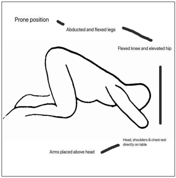 |
Fig. (1). Diagram showing knee-chest decubitus position. |
For the Free activities group (Group B): Pregnant women were allowed free activities like walking, or climbing stairs. Both groups of pregnant women were re-examined 5 minutes after the activity to check for a clear facial feature. If the image was still unclear, they were asked to continue the respective activity for 15 minutes and 30 minutes with intermittent Ultrasound imaging to check if the face was clear or still obstructed. If after 30 minutes of activity, the fetal face was unclear, then the female was asked to return the next day for re-imaging by 4D Ultrasound.
3. RESULTS
Knee-chest Decubitus position group (Group A): 5-minute post Knee-chest position, Four-dimensional Color Doppler ultrasound showed a clear fetal facial structure in 185 people; 15-minute post was observed in 86 people; 30 minutes post was observed in 77 people. For 12 people, a clear facial feature could not be obtained even after 30 minutes and they were asked to come back the consecutive day for re-examination.
Free activity group (Group B): 5-minute post free activity, Four-dimensional Color Doppler ultrasound showed a clear fetal facial structure in 72 people; 15 minute post was observed in 90 people; 30 minutes post was observed in 140 people. For 58 people, a clear facial feature could not be obtained even after 30 minutes and they were asked to come back the consecutive day for re-examination.
Re-examination for both the groups returning on the second day was successful.
Graph. (1) compares the success rates in both Groups utilizing the two different maneuvers using the following data.
 |
Graph. 1. Comparison of Success rates in two different maneuver groups. |
3.1. Data
Two groups of pregnant women after the intervention showed the success rate of facial comparison [n(%)] as follows:
Group A: Post 5,15,30,>30 minutes/1Day: 185 (51.4%), 86 (23.9%), 77 (21.4%), 12 (3.3%)
Group B: Post 5,15,30,>30minutes/1Day: 72 (20%), 90 (25%), 140 (38.9%), 58 (16.1%).
Figs. (2 to 4) shows the obscured and unobscured fetal facial features in both groups A and B.
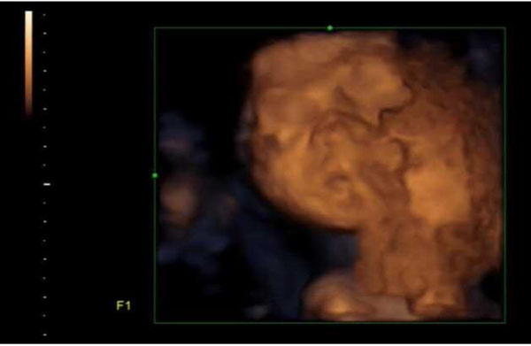 |
Fig. (2I). 4D Ultrasound Imaging of Fetal Face at the beginning of the test for a Group A Patient. Facial feature is unclear because its blocked by the hand and arms of the fetus. |
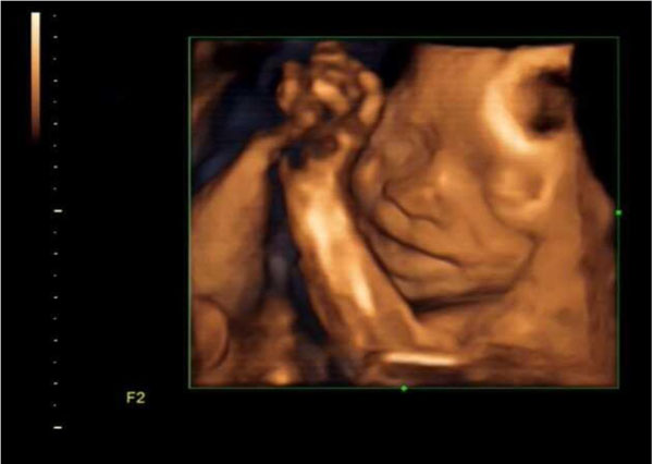 |
Fig. (2II). Successful 4D Ultrasound Imaging of Fetal Face with clear features Post 5 minutes of Knee-Chest Decubitus Positions in the same Group A patient as in Picture 1. [I]. |
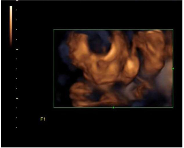 |
Fig. (3I). 4D Ultrasound Imaging of Fetal Face at the beginning of the test for a Group B Patient. Picture is unclear because face being blocked by artifacts and limbs. |
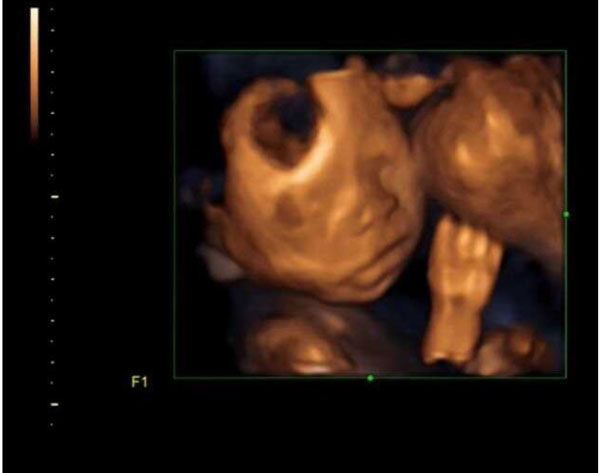 |
Fig. (3II). Successful 4D Ultrasound Imaging of Fetal Face with clear facial features Post 30 minutes of walking in that same Group B patient as in picture 2. [I]. |
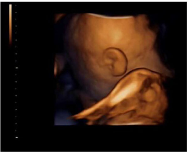 |
Fig. (4I). 4D Ultrasound Imaging of Fetal Face at the beginning of the test for a Group A Patient. |
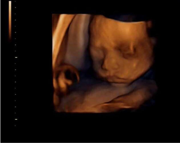 |
Fig. (4II). Successful 4D Ultrasound Imaging of Fetal Face with clear facial features post 5 minutes of Knee-Chest Decubitus Position for that same Group A Patient. |
4. DISCUSSION
Four-dimensional ultrasound imaging [2] of the fetus in pregnant women is highly acceptable these days because it enables pregnant women to see their baby in their own body. This is one of the most touching moments as it marks the successful establishment of the bond [5] between mother and child. So, many pregnant women are likely to choose this procedure. However, the four-dimensional imaging surface model needs a good sound window set off and an ideal fetal position in order to obtain three dimensional, intuitive and clear images. In a number of cases, some fetuses in the abdomen were prone or their faces were obstructed by the limbs, umbilical cord [8] or other obstructions. As a result, it is difficult to see the clear face of the fetus during ultrasound imaging. The traditional and widely used method [4], which is used to achieve an unobstructed view of the fetal face is to let the pregnant women move freely or climb stairs. The success rate, in that case, is found to be 20% in 5 minutes and 25% in 15 minutes. Nearly half of the pregnant women needed to walk for half an hour or more, which consumed a lot of time. So, physically and mentally exhausted pregnant women were often likely to give up the quest automatically. In contrast, the knee-chest decubitus position results in fully acceptable fetal facial clarity in a very short span of time. A 51.4% success rate is achieved in 5 minutes. This method besides being simple, safe and effective, is substantially more convenient for patients, clinics and hospitals and also is therefore worth promoting.
This is such an amazing discovery in the field of Fetal ultrasound [9], because it enables the mother to see her child’s face even before delivery and establishes a more human touch and connection developed with her. There are instances when the clear visualization of the fetal face would not be possible because of the face being obstructed with fetus’s other body parts. So it is very important to manipulate different positions in order to be able to obtain a clear face of the fetus.
Sometimes, even after a long duration of walking by the mother, it is still impossible to obtain a clear image of the fetal face. In that case, we ask them to lie down on their belly for some time, that enables us to get a clear picture.
This is not harmful to the pregnant mother as neither does this compromise any blood flow to the fetus, nor does this affect the pregnant mothers health condition [10].
The only kind of problem which might be encountered while examining few patients would be their unwillingness to lie down in the same position for more than 5 minutes or just an uneasy feeling while lying down in that position. This is not medically relevant but the patient’s wishes should be acknowledged. On the other hand, rather than waiting for almost 30 minutes or sometimes even till next day to get a clear view of the fetus, the knee-chest maneuver enables a mother to quickly get a clear and unobstructed view of her unborn child.
5. LIMITATIONS
Although Knee-chest decubitus position considerably decreases the total time required for a patient to achieve a 4D ultrasound image, there are minor limitations [11]. Some advanced stage pregnant patients would not be willing to lie down in this position given their inner discomfort and anxiety. Some patients might not agree to lie down for such a long time in the same position. Furthermore, allowing one patient to lie for 30 minutes in a Knee-chest decubitus position would be quite inefficient resulting in a prolonged waiting period for a large number of patients in the queue to get checked. Nevertheless, the present study indicates that the knee-chest procedure will be unsuccessful in a substantially smaller proportion of patients than the free activity strategy [12]. As benefit outweighs the risk, this maneuver can still be considered to cut down on valuable examination time and be used successfully worldwide.
CONCLUSION
As seen from the study, the knee-chest decubitus position compared to the free activity proved to be successful in the delineation of the fetal facial structure. In a larger proportion of patients for whom the first-time imaging was not able to provide a clear picture, knee-chest decubitus position for 5 minutes proved to be quite successful. If this maneuver is carried out, it will successfully reduce real time ultrasound imaging time and also reduce the disappointment and stress of the mother when not being able to view the fetuses face at the first instance.
ETHICS APPROVAL AND CONSENT TO PARTICIPATE
The study was approved by Basic research program and Science foundation of the Traditonal Chinese Medicine Hospital affiliated to Southwest Medical University.
HUMAN AND ANIMAL RIGHTS
No Animals were used for studies that are base of this research. The reported experiments were in accordance with the ethical standards of the committee responsible for human experimentation (institutional and national), and with the Helsinki Declaration of 1975, as revised in 2008.
CONSENT FOR PUBLICATION
Written and informed consents were obtained for the study from all the patients involved in the experiment.
CONFLICT OF INTEREST
The authors declare no conflict of interest, financial or otherwise.
ACKNOWLEDGEMENTS
This research is partially supported by the Basic Research Program and Science Foundation of The Affiliated Traditional Chinese Medicine Hospital of Southwest Medical University, China.
There is no conflict of interest and all the Pregnant Patients signed an informed consent before undergoing the 4D fetal ultrasound imaging.
